Home

poslucháč Veľká noc Reshoot iba1 marker voľný čas pohodlie chyžná

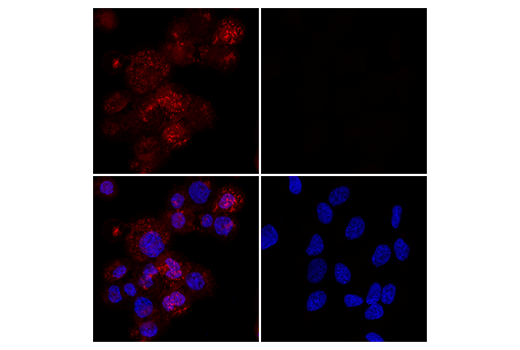
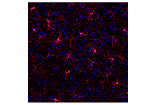
Iba1/AIF-1 (E4O4W) XP® Rabbit mAb (Alexa Fluor® 594 Conjugate) | Cell  Signaling Technology
Iba1/AIF-1 (E4O4W) XP® Rabbit mAb (Alexa Fluor® 594 Conjugate) | Cell Signaling Technology

Anti-AIF1 / Iba1 Monoclonal Antibody(Clone: AIF1/1909)

Fig 1 | PLOS Biology

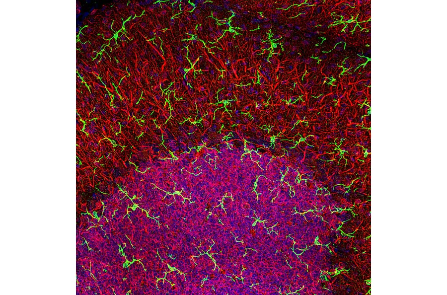
Expression of microglia marker (Iba1, green) by mouse brain cells... |  Download Scientific Diagram
Expression of microglia marker (Iba1, green) by mouse brain cells... | Download Scientific Diagram

Co-expression of Iba-1 and M1/M2 polarization markers. Brain slices... |  Download Scientific Diagram
Co-expression of Iba-1 and M1/M2 polarization markers. Brain slices... | Download Scientific Diagram

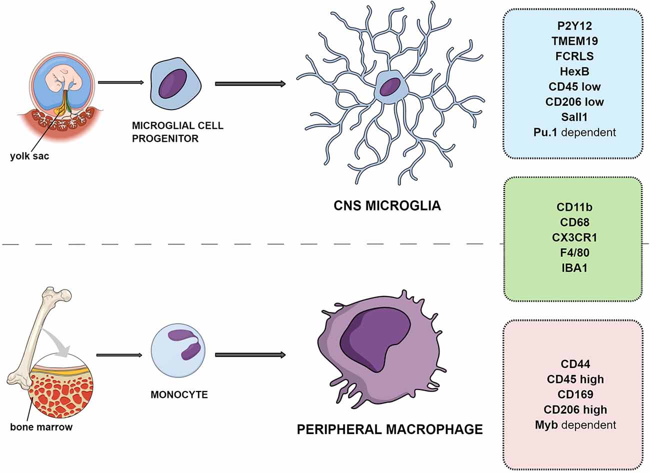
Frontiers | Overview of General and Discriminating Markers of Differential  Microglia Phenotypes
Frontiers | Overview of General and Discriminating Markers of Differential Microglia Phenotypes

Cells | Free Full-Text | Beyond Activation: Characterizing Microglial  Functional Phenotypes
Cells | Free Full-Text | Beyond Activation: Characterizing Microglial Functional Phenotypes

iReal Biotechnology, Inc. | iReal Biotechnology, Inc.

Neuroinflammation Marker (BDNF, ICAM1, TREM2, GFAP, TNF alpha, Iba1)  Antibody Panel - Human (ab263462)
Neuroinflammation Marker (BDNF, ICAM1, TREM2, GFAP, TNF alpha, Iba1) Antibody Panel - Human (ab263462)
Iba1/AIF-1 (E4O4W) XP® Rabbit mAb (Alexa Fluor® 594 Conjugate) | Cell  Signaling Technology
Iba1/AIF-1 (E4O4W) XP® Rabbit mAb (Alexa Fluor® 594 Conjugate) | Cell Signaling Technology

A Guide to Microglia Markers | Biocompare: The Buyer's Guide for Life  Scientists
A Guide to Microglia Markers | Biocompare: The Buyer's Guide for Life Scientists

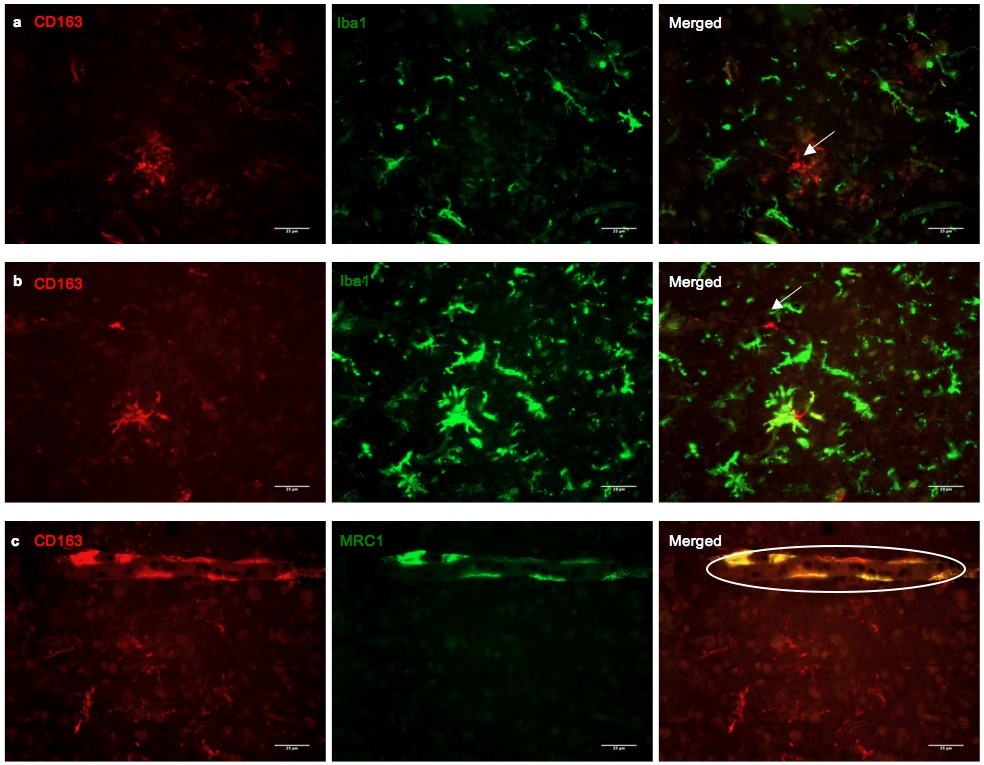
Phenotypic profile of alternative activation marker CD163 is different in  Alzheimer's and Parkinson's disease | Acta Neuropathologica Communications  | Full Text
Phenotypic profile of alternative activation marker CD163 is different in Alzheimer's and Parkinson's disease | Acta Neuropathologica Communications | Full Text

Quantitative immunohistochemical analysis of myeloid cell marker expression  in human cortex captures microglia heterogeneity with anatomical context |  Scientific Reports
Quantitative immunohistochemical analysis of myeloid cell marker expression in human cortex captures microglia heterogeneity with anatomical context | Scientific Reports

GeneTex Introduces Recombinant Monoclonal Iba1 Antibody | GeneTex

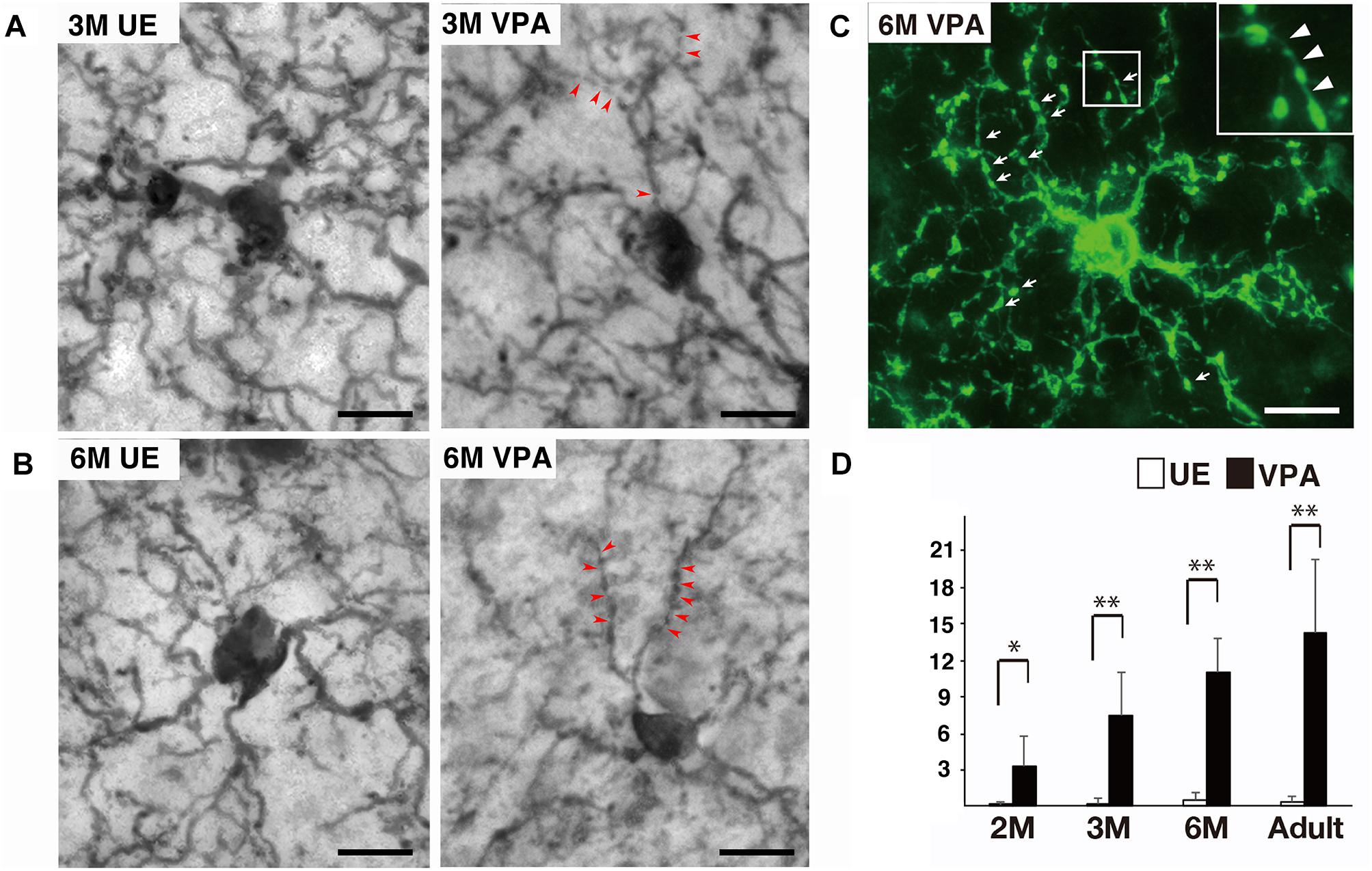
Frontiers | Segmented Iba1-Positive Processes of Microglia in Autism Model  Marmosets
Frontiers | Segmented Iba1-Positive Processes of Microglia in Autism Model Marmosets

Microglia markers | Abcam

Anti-Iba1 Antibody (A104332) | Antibodies.com

Iba1/AIF-1 (E4O4W) XP® Rabbit mAb (Alexa Fluor® 555 Conjugate) | Cell  Signaling Technology
Iba1/AIF-1 (E4O4W) XP® Rabbit mAb (Alexa Fluor® 555 Conjugate) | Cell Signaling Technology

Microglia Markers
Microglia Markers

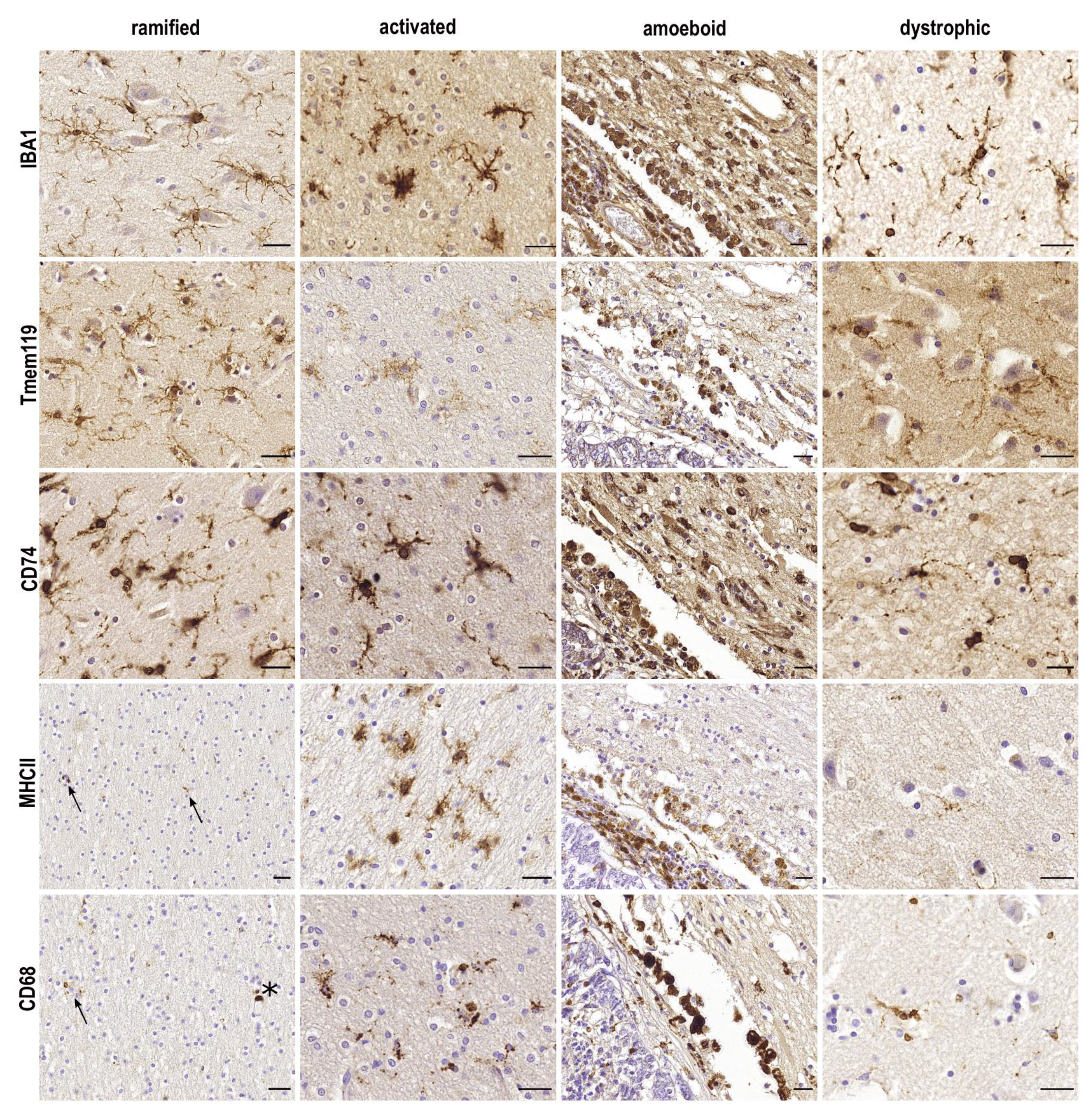
Staining of HLA-DR, Iba1 and CD68 in human microglia reveals partially  overlapping expression depending on cellular morphology and pathology -  ScienceDirect
Staining of HLA-DR, Iba1 and CD68 in human microglia reveals partially overlapping expression depending on cellular morphology and pathology - ScienceDirect

Distribution of microglia/macrophage marker, Iba1, and inflammatory... |  Download Scientific Diagram
Distribution of microglia/macrophage marker, Iba1, and inflammatory... | Download Scientific Diagram

Quantitative immunohistochemical analysis of myeloid cell marker expression  in human cortex captures microglia heterogeneity with anatomical context |  Scientific Reports
Quantitative immunohistochemical analysis of myeloid cell marker expression in human cortex captures microglia heterogeneity with anatomical context | Scientific Reports

Blood-derived GFP+ monocytes that infiltrate the brain parenchyma following  HSV-1 infection express the microglia marker Iba1.
Blood-derived GFP+ monocytes that infiltrate the brain parenchyma following HSV-1 infection express the microglia marker Iba1.
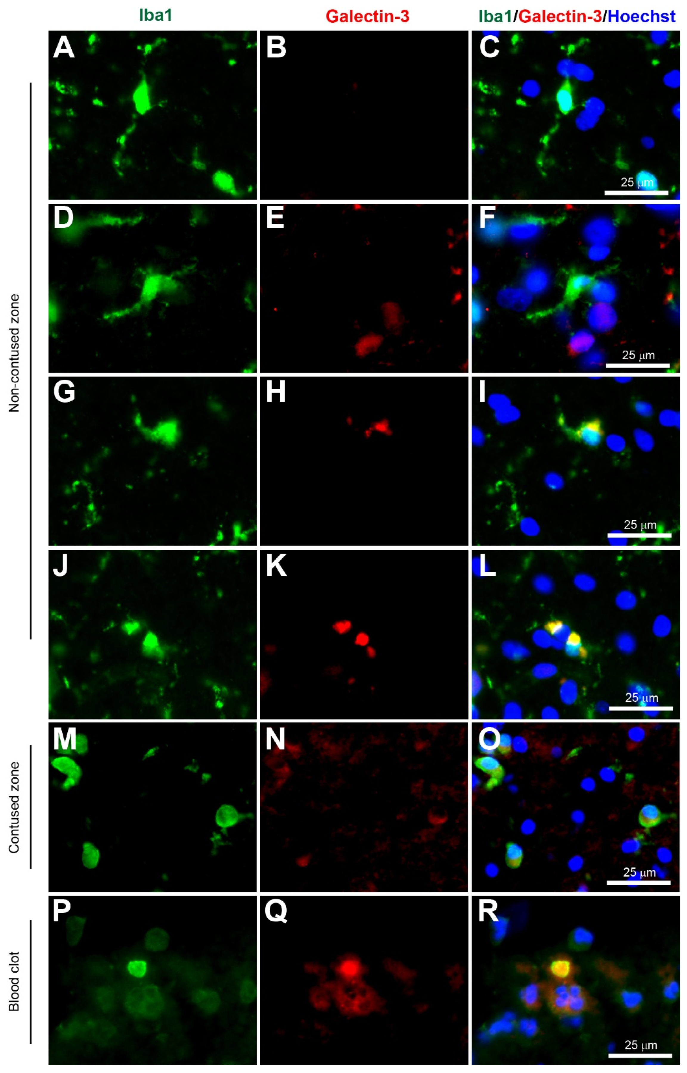
Biomolecules | Free Full-Text | Advances in Visualizing Microglial Cells in  Human Central Nervous System Tissue
Biomolecules | Free Full-Text | Advances in Visualizing Microglial Cells in Human Central Nervous System Tissue