Home

posol menej všeobecný magnetite thin section hotel kníhkupectvo lyžice

Photomicrograph Tuesday: Magnetite & hematite | Life in Plane Light

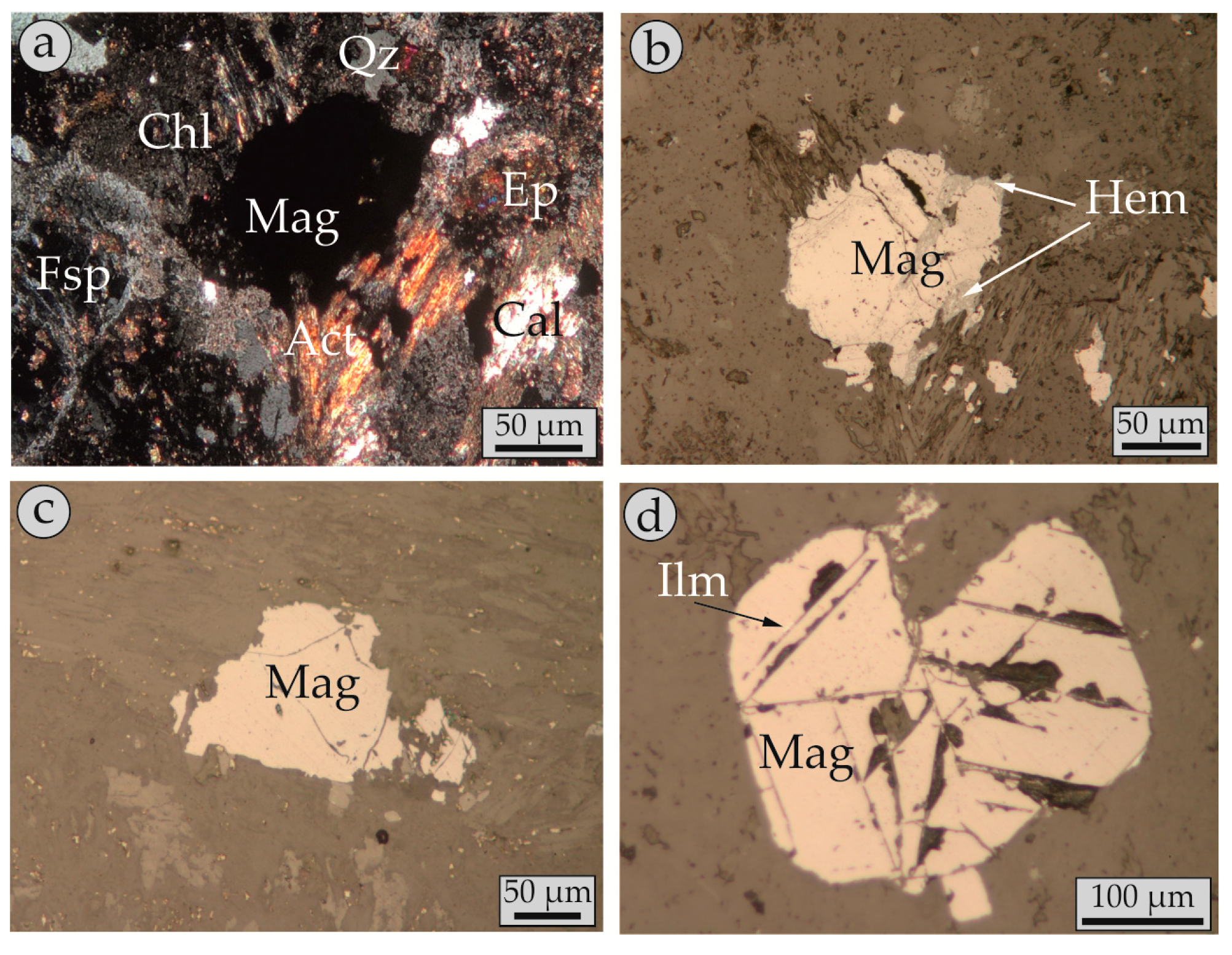
Minerals | Free Full-Text | Trace Elements in Magnetite from the Pagoni  Rachi Porphyry Prospect, NE Greece: Implications for Ore Genesis and  Exploration
Minerals | Free Full-Text | Trace Elements in Magnetite from the Pagoni Rachi Porphyry Prospect, NE Greece: Implications for Ore Genesis and Exploration

Magnetic characterisation of magnetite and hematite from the Blötberget  apatite – iron oxide deposits (Bergslagen), south-central Sweden
Magnetic characterisation of magnetite and hematite from the Blötberget apatite – iron oxide deposits (Bergslagen), south-central Sweden

A: Plane polarized light (PPL) image of thin section of chert... | Download  Scientific Diagram
A: Plane polarized light (PPL) image of thin section of chert... | Download Scientific Diagram

Thin section in PPL (plane-polarized light). R-82-6 sample,... | Download  Scientific Diagram
Thin section in PPL (plane-polarized light). R-82-6 sample,... | Download Scientific Diagram

Hematite | NOVA Mineralogy

ALEX STREKEISEN-Magnetite-

O3. Magnetite Gabbro

Magnetite composition in Ni-Cu-PGE deposits worldwide: application to  mineral exploration - ScienceDirect
Magnetite composition in Ni-Cu-PGE deposits worldwide: application to mineral exploration - ScienceDirect

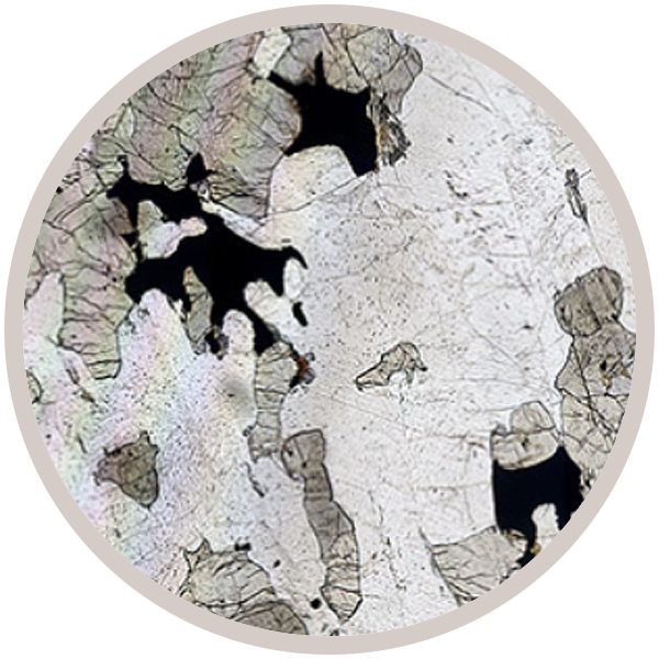
2.6 Properties Under Plane Polarized Light – Introduction to Petrology

Figure F82. Inclusions of pyrrhotite and magnetite in pyrite (Sample  193-1188F-11G-1 [Piece 9, 108-111 cm] in reflected light; width of view =  0.275 mm. Photomicrograph ID# 1188F_23; thin section 74).
Figure F82. Inclusions of pyrrhotite and magnetite in pyrite (Sample 193-1188F-11G-1 [Piece 9, 108-111 cm] in reflected light; width of view = 0.275 mm. Photomicrograph ID# 1188F_23; thin section 74).

Proc. IODP, 345, Hole U1415P

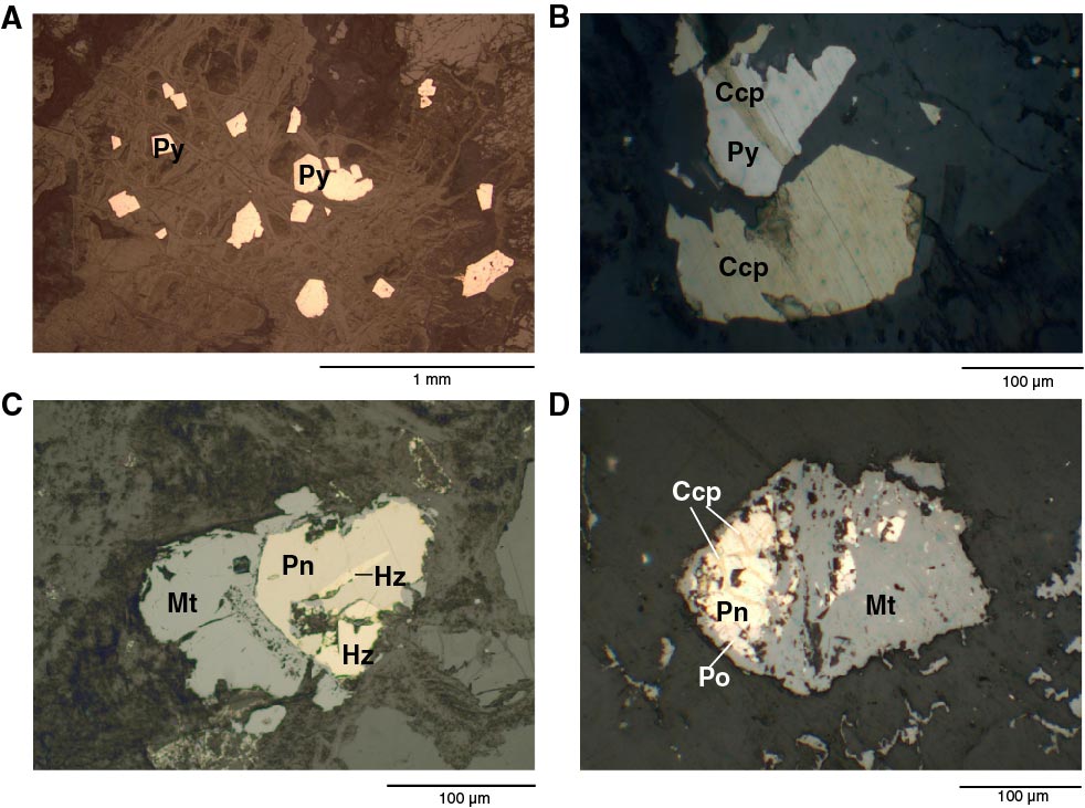
a. Photomicrographs of magnetite ore with pyrite, and chalcopyrite in... |  Download Scientific Diagram
a. Photomicrographs of magnetite ore with pyrite, and chalcopyrite in... | Download Scientific Diagram

2.6 Properties Under Plane Polarized Light – Introduction to Petrology

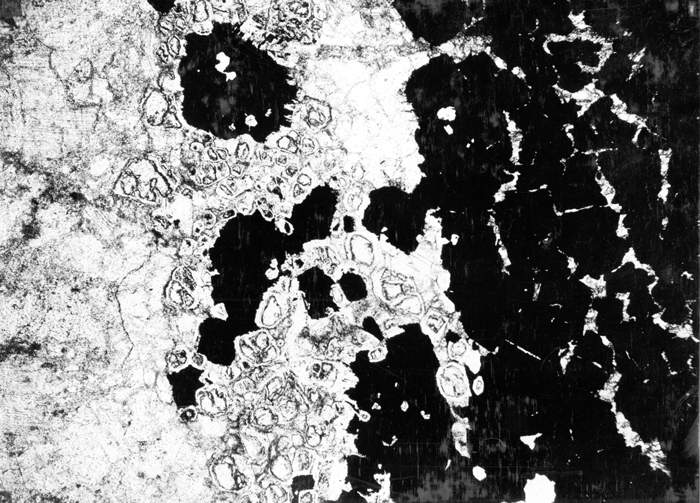
Thin section across margin of magnetite bodies shown in Calki... (1) -  Photo Gallery 4 -
Thin section across margin of magnetite bodies shown in Calki... (1) - Photo Gallery 4 -

Photographs of representative thin sections showing textures of (A, B,... |  Download Scientific Diagram
Photographs of representative thin sections showing textures of (A, B,... | Download Scientific Diagram
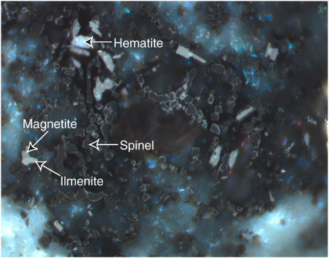
Iron-Titanium Oxides - Geology North

Thin section in PPL (plane-polarized light or II -as symbol). R-82... |  Download Scientific Diagram
Thin section in PPL (plane-polarized light or II -as symbol). R-82... | Download Scientific Diagram

Figure F90. Spinel (dark gray), magnetite (medium gray), ilmenite (light  gray), and hematite (white). Spinel has a thin coating of magnetite.  Magnetite contains inclusions of hematite. Magnetite-ilmenite intergrowth  on the left side
Figure F90. Spinel (dark gray), magnetite (medium gray), ilmenite (light gray), and hematite (white). Spinel has a thin coating of magnetite. Magnetite contains inclusions of hematite. Magnetite-ilmenite intergrowth on the left side

Magnetite exsolution in ilmenite from the Fe-Ti oxide gabbro in the Xinjie  intrusion (SW China) and sources of unusually strong remnant magnetization
Magnetite exsolution in ilmenite from the Fe-Ti oxide gabbro in the Xinjie intrusion (SW China) and sources of unusually strong remnant magnetization

Hematite | NOVA Mineralogy

Figure F83. Inclusion of hematite (medium gray) with a remnant of magnetite  (dark gray) in pyrite (white) (Sample 193-1188F-35Z-1 [Piece 2D, 44-46 cm]  in reflected light; width of view = 0.14 mm.
Figure F83. Inclusion of hematite (medium gray) with a remnant of magnetite (dark gray) in pyrite (white) (Sample 193-1188F-35Z-1 [Piece 2D, 44-46 cm] in reflected light; width of view = 0.14 mm.